Tendonitis Surgical Photos
Before and After Release of the Retinaculum

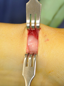
Before Tendonitis Relief Surgery
The thick extensor retinaculum (sheath) that needs released as it pinches tendons.

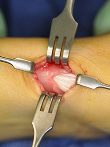
During Surgery
A partial release of the retinaculum.

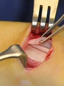
After Surgery
Complete release of the retinaculum. Note how thick this abnormal band is. It should be thin like cellophane.


